


Osteochondrosis is a chronic disease that is symptomatically expressed by dystrophic disorders in the articular cartilage. Osteochondrosis of the spine most often occurs when changes in intervertebral discs and herniated discs occur. Depending on the localization, cervical, thoracic and lumbar osteochondrosis are distinguished. Osteochondrosis is often found in which all vertebral sala parts suffer. The pathology requires a consultation with a doctor and an integrated treatment approach.

Description
Like other chronic diseases, diseases of the spine quickly become "younger". When the pain in the back and the joints previously disturbed older people, patients between the ages of 18 and 30 are increasingly treating doctors today.
Scientists regard the directness of a person as a prerequisite for the development of this disease. Osteochondrosis is also facilitated by a longer sitting position. In addition, the articular cartilage with old elasticity and elasticity, thinly loses thin; Intervertebral discs lose moisture and shock absorption and are susceptible in the period of physical exertion.
Reasons
The causes of cervical osteochondrosis are in loads and under different circumstances affect the cervix region. Accordingly, the muscles in the neck will take off intensively, which compensates for this load, causing a cramp to occur and a violation of blood flow in this area.
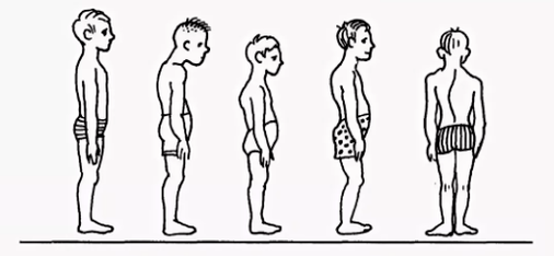
- Violation of attitude coliosis, bend, round back, kyphosis and other posture disorders, even if they are insignificant, cause a serious violation of the balance of the spine. As a result, the strain on the intervertebral discs is unevenly distributed, which provokes its deformation and increased wear. The vertebrae begin to bring yourself closer, which leads to violations of nerve processes. The cervical osteochondrosis develops pretty quickly. Similar consequences have violations of the attitude caused by a change in the natural position of the ribs.
- Muscle spasms of cramping reactions of the muscles of the back, the chest, press can cause individual parts of the body to be very tense. As a result, the general equilibrium position of the body is disturbed, which leads to a change in the position of the spine. Deformations can influence the area of the cervix region or other parts of the spine, which leads to osteochondrosis of the chest, cervix and loin parts.
- Violations of blood supply, since vertebrates have no direct connection to the circulatory system, you get a diet from surrounding tissues. The violation of the blood supply to the cervical spine means that the slices do not receive enough liquids for rehydratization (the restoration of the shape due to the absorption of moisture) and the renewal of the cartilage tissue. As a result, your wear is accelerated, a decrease in the distances between the vertebrae of the cervix region will decrease, which leads to osteochondrosis.
- Violation of innervation leads to a decrease in the sensitivity of nerve roots to pathological changes in its structure, whereby the shift and deformation of the vertebrae of the cervix region remain unnoticed by the patient. After all, the pain is missing due to sensitivity disorders.
- Diseases of internal organs are the wrong position of internal organs, the displacement and reduction of which lead to a violation of the general balance in the body due to various functional disorders. As a result, this influences the position of the spine column acutely - the cervix, the lumbar vertebrae are shifted and deformed, which leads to the corresponding types of osteochondrosis.
In general, the osteochondrosis of the cervix region develops due to the effects of disadvantageous external factors that violate the natural equilibrium position of the spine and other systems of the human body.
Diagnosis
The diagnosis of cervical osteochondrosis begins with the collection of all the necessary information about the patient. The specialist asks about symptoms that disturb the person is interested in his professional activities and how he spends his weekend. An important point is the presence of osteochondrosis among parents, grandparents, as this is an inheritance disease.
Then the doctor goes straight to the patient's visual examination. He studies the cervix compartment and his back to the curvature of the posture and gropes the cervix region. This enables a specialist to evaluate the degree of development of the disease, since in advanced cases the palpation of the cervix region leads to sharp pain.
When examining, you should be careful:
- About the severity of the cervix lordosis;
- the height of the shoulders in the patient;
- The possibility of the asymmetry of the Supraclock areas;
- the possibility of the asymmetry of the neck (e. g. a consequence of the congenital pathology or a sharp muscle cramp);
- the condition of the muscles of the shoulder strap and the upper limbs (e. g. a muscle atrophy can indicate compression of the cervical spine);
- The location of the chin - the chin is normal if there is a medium line.
- The movement of the neck (bending injury, tipping the right and rotation and rotation).
Palpation is carried out in the initial position of the patient:
- lie on the back;
- lie on the stomach;
- Sit on a chair.
The examination of the movement volume is also carried out. It is carried out in the initial position of the patient sitting on a chair (to repair other spine).
Differentiate the following basic movements in the cervix region:
- Flexion;
- Extension;
- Hang to the right and left;
- Rotation.
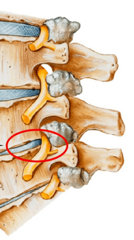
About half of the flexion and extension volume occurs between the back of the head, the vertebrae of C1 and C2. The rest of the movement is carried out due to the underlying vertebrae with a large number of movements in the C5-C7 vertebrae. The side groves are evenly distributed over all vertebrae.
In order to make a precise diagnosis, additional studies are prescribed:
- X -Ray of the cervix region. This method is appropriate in the early stages of the disease, but can be useless in advanced forms.
- CT (computer tomography). Allows you to see structural changes in the vertebrae, but with the help of this method it is impossible to determine the size of the hernia between the vertebrae.
- MRI. It is considered the most effective method for diagnosing cervical osteochondrosis. You can determine the size of the hernia between the windows and the degree of their development.
- The doctor can also prescribe a duplex scan with which you can determine a violation of normal blood circulation in the arteries.
Treatment
The treatment of cervical osteochondrosis is complex therapy that uses medication, gels and various physiotherapeutic measures. Therapeutic gymnastics and the massage of the problem area play an important role.
Medication
It is impossible to remove the consequences of degenerative-dystrophic changes in the spine without the use of medicinal products. The use of medicinal products in the treatment of osteochondrosis of the cervix region is one of the main points of the complex healing approach used by most traditional medicine specialists.
It was developed to solve several problems, including:
- Stop the pain symptom and remove the inflammatory process.
- Remove muscle cramp;
- stimulate the process of regeneration of cartilage and bone tissue cells;
- If you strengthen the body's protective properties, increase immunity;
- Improve the general condition by eliminating other symptoms that affect recovery.
Depending on these purposes, all medicines that are prescribed by a doctor who suffers from a cervical osteochondrosis can be divided into the following groups:
- Analgetics (non -steroid medication that relieve pain).
- Inhibitory anti -inflammatory (steroid) are hormonal medication that alleviates inflammatory phenomena and causing pain.
- Chondroprotectors contain medication that contain substances that replace the components of the cartilage tissue - chondroitin, hyaluronic acid).
- Musorelaxants. These are drugs that relax the muscle tone. They are used as aids in the operation and orthopedics to stop pain. Such drugs are administered by parentels and therefore always under the supervision of a doctor.
- Vitamins. In the case of osteochondrosis of the cervix region, vitamins are prescribed, which benefits the peripheral nervous system and the conductivity is improved. Water -soluble vitamins: B1, B6, B12, fat -soluble vitamins: A, C, D, E. In recent years, combined medication that contains both pain relievers and vitamin components more often.
- Ointments and gels for external use.

Physiotherapy
The main objective of physiotherapy treatment is to stimulate the regeneration process in the body and to eliminate pain. The most popular methods in the treatment of cervical chondrosis are the following:
- Ultrasonic. Physiuming with ultrasound waves is used to relieve serious pain manifestations and inflammatory reaction. Ultrasound is massaged by the neck tissue, according to which metabolism is activated.
- Vibration massage. The influence on the pain area during the vibration massage is through mechanical vibration movements. A strip vibror is usually used for the proper behavior of this physiotherapy.
- Electrophoresis. This method for carrying out the physiotherapeutic supply of cervical osteochondrosis is carried out using dianamic and modulated currents as well as electrical fields when the body is administered into the tissue of medicinal products. The electrophoresis perfectly relieves spasmodic syndromes and eliminates pain in inflamed muscles.
- Magnetotherapy. The essence of magnetotherapy as a physicalization in osteochondrosis of the cervix region is explained by the use of constants or variables of fields with a magnet, a frequency of different sizes. This method can help the patient remove pain and stop the inflammatory process in the stove. The procedure is often carried out at home after a special magnetographer has been recorded.
- Dutoenzor therapy. Currently there is a fairly popular physiotherapy method that exists with the stretching of the spine column under the mass of the body. In order to carry out such a procedure, a specially arranged mattress is required, which has inclined ribs and changes the location under the weight of your own body. The muscle tone is normalized, which leads to your relaxation.
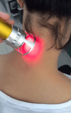
- Laser therapy. The laser has a complex influence on the focus of the inflammation and activates biological processes in the tissue of the nervous system. In this way you can have a positive effect from the treatment. A complex effect on the body is in anti -inflammatory, analgesic and wound healing effect. A laser treatment process should not exceed 15 minutes. This is the optimal time of the sectoral exposure of the Helik-Neon laser in the affected areas. In this case, the duration of the laser should not exceed 2 minutes if a pain.
- Balneotherapy. The advantages of mineral water have been known for a long time, the balneotherapy is based. The procedure implies the active use of water resources in the treatment of osteochondrosis. In addition to the introduction of baths, different types of souls and active swimming in the pool, the therapy includes the use of the therapeutic sludge to painful body areas. The therapeutic effect is achieved by the simultaneous effect of chemically active substances that are contained in water at different temperature conditions. With the technology you can stop pain syndrome by improving local microcirculation in the tissues.
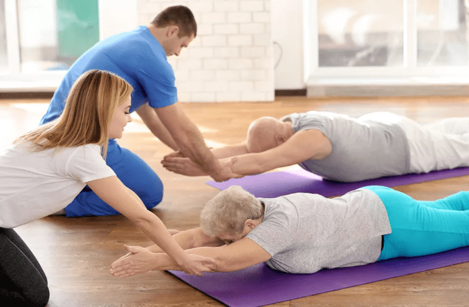
Training therapy
It should be remembered that training therapy is not carried out when signs of tightening begin: pain. According to the LFK complex, you can strengthen yourself and cause inconvenience.
There are a number of general recommendations for fees:
- Sports lessons should take place in the interior with good ventilation, an excellent option on the street.
- The classes are only carried out during the relief of the disease (if there are no symptoms).
- Clothing in training therapy should be broad, not embarrassing movements and breathing.
- All movements are smooth, the amplitude and the number of repetitions are gradually increasing.
- When the pain begins, you should stop the lesson immediately.
- Passes the classes ahead and end the measurements of pressure and pulse. If these indicators differ normally, the load should be reduced.
- It is advisable to listen to your breathing throughout the lesson, this will increase efficiency. All stretching exercises are carried out when exhaling.
- It is very important to gradually increase the load and the number of repetitions. This reduces the risk of injuries and prevents revisions.
- Exercises are important to carry out regularly so that you can achieve a quick result.
- Before you start independent classes, you have to consult a doctor and agree with a number of exercises.
Recommended exercises in the starting position that lie on the stomach:
- The head is on the forehead, hands on the back of the head, elbows parallel to the floor. Lift your head with your hands from the ground, keep this position on 4 accounts, lower and relax. Repeat 2-4 times.
- The head is in the chin, palms under the chin. Time in time you stretch your arms forward, two - except on the sides, three - forward, four - the starting position. Repeat 2-4 times.
- Hands stretched forward. Swimming in the "rabbit" style, repeated 4-8 times.
- Hand surfaces under the chin, emphasis on the palm of your forehead. Alternatively, remove the heel of the buttocks. Repeat 4-8 times.
Recommended exercises in the starting position, which is on the page (right, then left):
- The right hand is extended, the right ear lies on it, lift your right hand with your head, keep the position on 4 accounts, lower and relax. Repeat 2-4 times.
- The left hand rests on the floor in front of the chest, the left leg makes the fly movements back and forth. Repeat 6-8 times.
- The left hand along the body increases the left hand upwards, lower, lower. Repeat 2-4 times.
- The left hand on the thigh. If you pull both knees up to your chest when exhaling and smooth your legs with inspiration. Repeat the exercises 2-4 times.